In my newsletters, I often report on estrogen’s actions, especially estradiol. In the newsletter, Estrogen helps heal Traumatic Brain Injury, Ialso highlighted the role of the estrogen synthetase enzyme, aromatase. This enzyme is found in the vasculature, and may be involved in estrogen’s vasodilation effect.
Its redox reaction is thought to generate nitric oxide (NO), inducing smooth muscle relaxation.
Estrogen receptor signaling in the vascular endothelium. Membrane-bound ER-α co-localizes with calveolin-1 and signals through phosphatidylinositol 3-kinase (PI3K) and protein kinase B (Akt) as well as mitogen activated protein kinase (MAPK) to phosphorylate eNOS, triggering an increase in NO. ER-α signaling also increases intracellular calcium through phospholipase-C/inositol 14,5-triphosphate pathway which then subsequently binds calmodulin and activates eNOS. Membrane-bound ER-α can also activate eNOS in response to flow. GPER transactivates epithelial growth factor receptor (EGFR), and signals via ERK1/2/PI3K/Akt to activate eNOS). GPER upregulates calcium through the inositol 14,5-triphosphate and ryanodine receptors on the endoplasmic reticulum as well as transient receptor potential channel on the plasma membrane which triggers calcium/calmodulin signaling to promote eNOS phosphorylation. GPER signaling also increases expression of transcription factor Kruppel-like-factor 2 (KLF-2) , which subsequently increases eNOS expression and glycolytic pathways. Estrogen increases activity of sphingosine kinase, S1PR1, and S1P transporters, which increases NO production. Nuclear estrogen receptors upregulate antisenescent proteins, increase expression of eNOS, and enhance neutral sphingomyelinase /sphingolipid signaling. Image created using Biorender.com and published with permission. - G SenthilKumar et al.
But now new research implicates involvement of an ion channel.
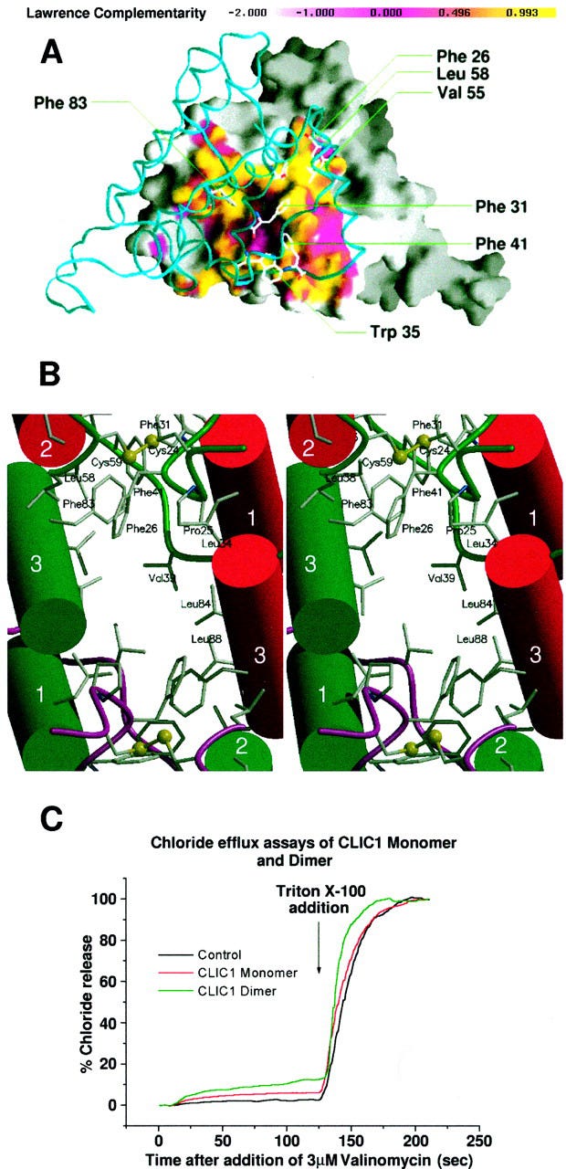
The dimer interface of oxidized CLIC1 plus channel activity. A, molecular surface of the A subunit colored by Lawrence shape complementarity, S(x), of the interface. A backbone representation of the B subunit is displayed with residues involved in the aromatic columns labeled. The overall value for S(x) of 0.720 is comparable with oligomeric protein interfaces. The tightly packed aromatic region of the interface (yellow) corresponds to S(x) values close to unity, and it encloses the loosely packed inner hydrophobic region with low S(x) values (pink). The figure was made using GRASP. B shows a stereo view of the dimer interface. At the top and bottom of the figure are the tightly packed columns of aromatic residues, whereas there is an aliphatic cavity in the center. Helices shown as per Fig. 2.C, valinomycin-dependent chloride efflux from liposomes: black line, control; red line, CLIC1 monomer; green line, dimer. After 120 s 0.1% Triton X-100 was added to release remaining intravesicular chloride. - DR Littler, et al.
BioMedWorks’ Newsletter is a reader-supported publication. To receive new posts and support my work, consider becoming a free or paid subscriber.
Keep reading with a 7-day free trial
Subscribe to BioMedWorks’ Newsletter to keep reading this post and get 7 days of free access to the full post archives.